Volume 04:

Episodes 076 - present

Types of shock:

☙ Cardiogenic - mechanical pump failure - infarcts, acute valve rupture dysrhythmias, myocarditis, hypoxemia, or poisoning events
☙ Obstructive shock - something is physically obstructing the system - pulmonary emboli, tension pneumothorax, cardiac tamponade, and acute RV infarct.
       - Third trimester pregnancy lying supine and compressing inferior vena cava causes obstructive shock - place a wedge under right hip and tilt baby off the midline.
☙ Hypovolemic shock - blood or fluid removed from body - a pool of blood, vomit, sweat, or severe third spacing.
☙ Distributive shock - fluid trapped in the vessels in the periphery unable to circulate due to vasopalgia - spinal cord injuries, sepsis, toxic causes.

 Four physiological variables associated with shock
#1 Preload - blood upstream from the heart waiting in the SVC and the IVC to enter the right atrium. Insufficient fluid decreases cardiac output and not fill completely. Ongoing supply of returning blood is critical in maintaining circulation.
       - resuscitation a woman with a gravida appearing uterus should be rolled to the left with a wedge
#2 After Load - resistance the heart must push against so for blood to exit the heart. If blood pressure is 120/80 it means the left ventricle must overcome 120mm/Hg of resistance. The higher the diastolic the greater resistance the heart must work to pump blood. Lower diastolic = less resistance/work.
After load important consideration: brain, kidneys, and pancreas all need a certain amount of blood pressure to be perfused and for their cells to not die
- Excess after load means the heart is working harder without physiologic benefit and the harder the heart works increases need for oxygen and glucose
- Demand ischemia - heart supply of oxygen is restricted due to pathology - coronary artery disease, profound anemia, tachycardia this creases a mismatch where the heart consumes more oxygen and glucose than can provide to itself.
Treatment:
       - increase the supply ex: giving oxygen.
       - reduce the amount of work the heart is doing. Reduce after load to minimum acceptable limits.
#3 Heart Rate - represents complete a cardiac cycle - relax and refill and to squeeze an eject. In physiological terms control of heart rate means control of optimal filling time. Frank Starling principle - filling time and filling volume equates to optimal efficiency of each beat.
#4 Contractility - Frank Starling curve - how myocytes have a different contractile strength and efficiency depending on how stretched out they are. The more the left ventricle fills with blood during diastole = more stretch towards their full optimal length. Systole achieves a greater ejection fraction - greater amount of blood from the ventricle makes it out through the aortic valve vs staying in the ventricle during the squeeze. Optimal filling is a major determinant for contractility or the efficiency of every heartbeat. Contractility through optimal filling highly influenced by preload as well as by filling time (heart rate)
Targets for four variables - mirror normal resting physiology for each of these variables.
Preload – want patients hydrated not flooded.
       - If intravascular volume low give IV fluids or blood
       - If blood circulating volume is missing or can’t increase blood returning to the heart ie. distributive shock type picture at the same time
       - if patient is overloaded with edema or crackles through lungs use diuretics to make the heart more efficient
Management of preload is not to blindly give 2 liters of crystalloid. Use clinical judgment to assess intravascular volume and total body water.
Check fluid status and preload assessment of IVC ultrasound and interstitial lung ultrasound and basic echo and other sophisticated scans.
afterload - comes down to map. simple calculation - weighted average of systolic and diastolic blood pressure readings rather than a simple average.

MAP = two parts diastolic plus one-part systolic blood pressure divided by three.
- Ex. BP of 90/62 is 60 + 60 + 90 = 210/3 = MAP is 70 (BP machines auto calculate the map)
Keep 3 potential MAP targets based on estimate of adult patients risk factors:
MAP targets for baseline healthy = 55.
MAP for cardiovascular disease = 65.
MAP for suspected closed head injury = 70

Ex of patients.
Brenda 40 yo female healthy with blunt trauma from an MVA. MAP of 55.
Bill 65 yo accountant with similar MVA appears to have a healthy baseline concerned for harden arteries and cholesterol plaques. MAP of 65
Roxanne 40 yo healthy has a blunt trauma from a high-speed crash on Kawasaki ninja worried about closed head injury. MAP7 of 70.
Management of MAP targets usually concerned about how to increase to achieve that minimum target. In some cases, lowering map is important ie. cardiogenic shock.
Decreasing unnecessary after load means the heart in cardiogenic shock can improve output without as much energy - treating or avoiding demand ischemia.
 heart is working hard and becoming ischemic burning more energy than supplying.

How to increase MAP
1. Reverse underlying cause.
2. Use the 4 cardiac variables - ensuring adequate preloaded, heart rate, and contractility. Adding vasopressors with the correct balance a vasoconstrictive alpha adrenergic effects and beta adrenergic for adequate heart rate.

Keep an eye on map and titrate vasopressors accordingly. If BP increases with other treatment such as NS bolus or on own, maintain MAP as is unless patient becomes symptomatic.
- if patient symptomatic and need to decrease MAP treat the root problem. If pt in pain treat pain first decreasing sympathetic drive.
Heart Rate Targets – 60-80bpm
- low end of heart rate range – 60bpm
- heart rate less than 50 consider beta adrenergic
- heart rate greater than 80
- cardia tamponade – stroke volume reduced due to pressure on heart. cardiac output directly proportional to heart rate. treat the underlying problem and drain that fluid
contractility – optimized best by preload, after load, and heart rate.
If contractility still poor rule out other causes and consult with cardiology or other specialists
- MI
- Myocarditis
- Cardio toxic event
Bed side ECHO – practice frequently.

Interconnectedness of the variables -preload (central venous pressure), after load (map),
heart rate, contractility (stroke volume)
- stroke volume - blood moved through aortic valve with each heartbeat is proportional to the amount preload, as well as the filling time from heart rate.
- SV= HR x preload
- CO= HR x SV
- B/P = CO x Systemic Vascular Resistance (SVR)               


Dan Ostopowicz – family med in eastern Ontario working clinic and hospital setting with lab and x-ray 24/7 and diagnostic ultrasound 4 days a week 8-4.

Ultrasound education minimal during residency as experience in tertiary centre had it. Joined Rural ultrasound fellowship just finishing up. There are lots of resources available at present.

Prior to doing fellowship spent a lot of time and energy doing one day courses or reviewing online material without gaining more confidence.
Now uses ultrasound daily in different settings not previously used.

CASE 1
Male patient in 70-80s sent from family physician for worsening dyspnea, increased oxygen demands, and hypotension. Known history of heart failure, interstitial lung disease from amiodarone uses prior. Ultrasound used to look for plural effusions, B-lines, and cardiac function. In this case found large pericardial effusion and helped provide definitive treatment and send pt off to tertiary centre, rather than assume heart failure and diuresis patient.
Outcome was that patient had to wait a day for fluid to be drained as he was anticoagulated to make the procedure safer and had 1.5L of pericardial fluid removed.

CASE 2
Man in his 80s living off the grid for last 20-30 years. No running water, electricity, medical care. Patient was able to maintain himself with no issues prior to this event. Developed pain in leg so went and bought himself a cane with no help. Bought a walker with no help and resulted in a fall. Admitted with query gout as physical exam was unremarkable and had an elevated uric acid in blood work. Definitive diagnosis for gout is with joint aspiration with ultrasound guidance. Aspiration found purulent discharge and changed concern to septic joint. Patient sent to orthopedic surgeon where they drained ankle.

CASE 3
Female in her 80s with heart failure with reduced ejection fraction and admitted for heart failure exacerbation. Started on diuresis with daily weights. Patient developed abdominal pain – labs ordered – CBC, liver enzymes, lipase, VBG, lactate. Lactate came back elevated at 7. Used ultrasound to look for any biliary or pancreatic pathology, bowel obstruction, and looked at heart that showed reduced ejection fraction with significant valve regurgitation in tricuspid and mitral valves. Consulted with intensivist and shared ultrasound images and determined that patient was likely suffering from a low flow state due to ejection fraction being so poor and systolic function and regurgitation issues causing backup causing a diffuse hypo perfused state for patient. This led to patient being able to remain in community with repeat exams and ongoing treatment.

Ultrasound helps to understand pathology and open discussion with specialist and allow for definitive treatment plans.
Change in practice going forward is that ultrasound is now used daily in all settings from clinic to hospital rounds to ED. Helps provide clarity and prevents having to send people out of town for other tests.


Advise:
Don’t hesitate to ask for help
Keep learning and being curious
Put the probe on everything to get improve confidence, skills, and images.


Case
EMS called to 67yo male with 100pack/year smoking history. Avoiding health care. SPO2 60% on RA. Living in a hotel with no family support.  

Vitals on arrival 
☙ BP - 130/70 ☙ Pulse - 110 ☙ Temp - 36.2 ☙ Resp - 32 ☙ SPO2 - 60 on RA improved to 93% on simple face mask
After arrival 
☙ BP - 130/70 ☙ Pulse - 120 ☙ Temp - 36.2 ☙ Resp - 26 ☙ SPO2 - 93%

Limited air entry and subtle wheezes which EMS stated was improved from initial presentation after giving back-to-back salbutamol MDI. Patient moved to resus bay and put on NC to try to wean off O2. Monitors applied. IV access not obtained yet. 

While talking to pt he is head bobbing and falling asleep and hard to answer yes/no questions. 
Altered level of conscious pneumonic – DIMES
D – drugs
I – Infection
M – Metabolic
E – Environmental 
S – Structural
No pinpoint pupils, bystanders states other than smoking patient clean. No coughing no outwards signs of infection. Metabolic is long differential – EMS ruled out BGL concerns with BGL being 5.7. no hyper/hypothermia can also do co-oxmitry which also reads amount of carbon monoxide this site had one and it read at 6 (consistent with smoker) and below 10 which is concerning level. 

LABS – CBC, Lytes, Creatinine, VBG, LFT and lactate. Can also do CRP and TSH. 

☙ Initial diagnosis – respiratory failure – even with improvement of SPO2 highly suspicious of respiratory acidosis. Excess CO2 in blood is causing narcosis effect in brain stem. This is NOT an oxygenation failure in lungs this is a ventilation failure. 

☙ RECAP of normal physiology – physiology of oxygenation carried by hemoglobin is independent of physiology of ventilation (elimination of CO2). CO2+H2O->H+HCO3 that then gets dissolved into blood and released through alveolar membrane as CO2.

☙ Oxygenation and ventilation are two separate processes that share the lungs.
In this patents case oxygenation was fixed but ventilation was not. When you put a patient on O2 (in this case 5x room air at 21%) you maintain healthy SPO2. If you increase FiO2 you can maintain SPO2 with lower ventilation rates simply with amount of oxygen available in tidal volume. Average healthy adult has 5L per min tidal volume. 
Ventilation is not affected by changing FiO2. How do you decrease the ETCO2 in lungs? Answer is NOT to turn up the oxygen, rather CO2 elimination is proportional only to minute ventilation (MV) = Respiratory rate x tidal volume. In health adult MV = 500ml x 10resp/min = 5L gas exchange per min.

☙ Patient resp failure due to combined oxygenation and ventilation failure. Corrected oxygenation failure (with O2 and salbutamol increasing FiO2 by about 5) but ventilation failure more subtle to detect and did not correct. Need blood gas – that takes time to obtain or ETCO2 that is quantitative in a closed loop (typically intubated patient) it is hard to miss ventilatory failure.

☙ Patient condition at this point – patient resp around 18/min with approx. 200ml tidal volume (need to be on closed loop breathing to get accurate tidal volume). Gas exchange only takes place in alveoli. Bronchi, bronchioles, trachea and pharynx do not have gas exchange making up approx. 150ml in average adult of anatomical dead space. So effective gas exchange in this patient is only 50ml. MV – 50mlx20 = 1L of gas exchange. This is enough to keep SPO2 high enough, but CO2 is building up. This results in increased H+ in blood leading to acidosis. 

VBG – pH - 6.8 – causes brainstem inhibition or narcosis. PCO2 – 110 (normal is 40). Bicarb of 40 (normal is 28) – Ventilatory failure in the presence of falsely reassuring oxygenation. This is profound respiratory acidosis with metabolic compensation (due to bicarb) meaning this is not an acute event. Kidneys have been working overtime probably for weeks to combat acidosis. 

Silent lungs need aggressive bronchodilator and steroid tx. IV started and dose of methylprednisolone given 125mg. several nebulizers of Combivent (salbutamol & ipratropium bromide). Salbutamol IV – 2.5ml of neb fluid diluted with NS so you can give 250mcg (1/10th neb concentration). Helped improve lung capacity with increased air entry heard throughout. Could hear proper wheezes. When silent lungs you need to open the airways enough to be able to hear the wheezes. 

Patient is still hypoventilating due to pH and brainstem being suppressed. Trying to use a BVM and time it with pt own resp is difficult. BiPAP – used (consulted respiratory therapist to set up) used FiO2 -100% and inspiratory support of 12cm of H2O and PEEP of 5cm. This increases pt own tidal volume with each breath by augmenting inspirations and the PEEP helps keep alveoli from collapsing – preventing recurrent atelectasis with each breath. 
Patient condition improved. SPO2 increased to high 90s and repeat VBG showed improvement in pH and CO2

CBC – WBC – 11.5 – rest of blood work came back ok. 
Dose of piptaz to cover possibility of infection. 
Bedside US to rule out plural effusion, pulmonary edema, pneumothorax. 
Chest x-ray – requested by accepting hospital

Unable to wean off bi-pap – policy states that if a pt requires greater than 1-2 hours of support they need to be transported for safety. This requires 1:1 monitoring. Transported to ICU. 

Take home points
1. Respiratory rate and SPO2 value do not paint whole resp picture. VBG is best option for quantifying CO2 elimination status. The PA component of ABG is not necessary.
2. Try to anticipate underlying pathophysiology and avoid delaying treatment when treatment is not that risky. 
3. Be aggressive in reversing COPD exacerbations. Silent chests are scary – back to back MDI or nebs, low threshold for giving IV salbutamol are cornerstones in reversing. Also, low risk for giving steroids or antibiotics to these patients because these COPD patients (especially with and altered LOC) crash quickly. 


Career Path Choices with Capt Charles Kotulski

Disclaimer – Interview represents personal opinions and not those of the United States Army.

Background MD. US ARMY military physician- family medicine. Has completed EM fellowship prior to joining military

Recently returned from Korea after 1 year tour. Prior was in direct commission course followed by basic officer leadership course. Korea – served as family physician in largest overseas clinic servicing 12,000 patients, as well as operating as medical director for clinic. Typical military facility. Level 3-4 major trauma centre without the specialist care. 

Issues with running facility
☙ Complications of running the facility was the bureaucracy surrounding facility. Military for as much as it tried to be helpful, was it also hindered the facility. Lack of resources, not with Korea itself but the fact of being in the military setting and having to request everything from the US. If there is a shortage in the US, then base was last to receive supply.
       - Hypothetical Ex. Needing a box of band-aids, and needing to order through the US, when there was a pharmacy nearby that you could purchase Band-Aids but unable to do so. 

☙ Tours only 1 year long. When people start leaving, they are finally familiar with things, and then they leave starting over with new people who do not know facility. 

☙ Training of medical providers – not standard across the board and discrepancies noted.
       - Must be a US citizen if you want to work as a medical provider in the army
       - Training can be anywhere else, so long as it meets equivalency criteria as US training.
       - South Korean government service workers also allowed to work – different standards of care

☙ Transferring patents out of base most challenging.
       - Within the host nation or outside of country back to the US
       - During last 4-5 months there was a Korean doctor strike and transferring patients out to host nation was almost impossible.
       - General surgeons available to do some procedure but no ICU. Typically, didn’t keep severe septic shock patients especially if there was renal involvement as no nephrologist.
       - Psychiatric cases – sometimes challenging when it comes to a dependent or adolescent because psych ward in hospital could not accept these patients due to credentials and pediatric psychiatric.

Background prior to Army
Medical school Saba University – Family med residency in Washington State in Yakima. EM fellowship completed. Worked as a locum prior to starting military service.
**Mindset is important to dealing with resource limited settings.
       - Mindset that comes with experience – talking with colleagues about their experiences valuable
       - Having a mentor is helpful
Residency and fellowship training also helped as they were both in resource limited settings

ED in military sites are more like urgent care due to patient population – young healthy adults

Examples of critical cases
       - Septic shock – transferred out and managed with regular treatment but challenged with finding accepting hospital in South Korea.
       - Have neonatal service/OBGYN – no NICU. Any babies delivered under 34 weeks need transfer out. Had case of twins being delivered at 28 weeks, neonatal resuscitation. Higher occurrence than other medical/trauma emergencies.
o using people around you for their experience and knowledge very beneficial and goes along way.

ATLS, NRP, ACLS field training frequent. Every week half of the day was dedicated to sergeant training where people given time to catch up on admin or training or teaching.

Bedside ultrasound – no able to do as frequently due to patient population.
       - Try for couple times a week to preform bedside ultrasound. Practice on colleagues and have them practice on me.
Portable ultrasound – butterfly. Helpful to rule out RUQ pain for anything biliary. Cardiac patients valuable. SOB is a big issue in South Korea due to poor air quality index from pollution from China, North and South Korea. 6mo of soldiers being in country would develop sob due to pollution. Used ultrasound for many of those cases

Advise for any healthcare provider looking to work with own country military.
       - Talk to others who have done it before and get insight take with grain of salt. Things do change within large systems

Concerns with combat being in military – Doctors are more protected. Physician Assistants are sent more to active sites and physicians are more well protected.
RN’s – one of the most important and are everywhere in both active and non-active. Their specialty determines where they go.
All physicians are general medical officer GMO even if completed a specialty can be sent where they can be utilized most.

Civilian paramedics going into military – enter and enlisted and then then choose where they want to go.

Next steps - getting deployed to Support Unit – equivalent to level 3 trauma hospital in middle east for 6 months


094 - First Trimester Pain & Bleeding - Virginia Roberson

Video

Video Description


☙ Limited diagnostic tools in diagnosing embolic disease
D-dimer – high sensitivity poor specificity
       - Positive results generally require imaging
       - Challenging in rural centres
☙ Spectrum of embolic disease
       - Lungs filter out micro clots
       - When clots become excessive then we end up with pathological emboli
       - Trouble determining when it is a normal physiological function and changing to pathological condition
☙ Notoriety with patents – with google searches for symptoms and managing expectations of them worrying about life threatening issues. 

☙ Bedside POCUS
       - To rule out Deep Vein Thrombosis (DVT)
       - Can help with some Pulmonary Embolism (PE)
Weekend course to specify DVT ultrasound available
Online courses if familiar with basics of ultrasound, online has instructional videos to teach yourself.
       - Make sure you have a resource that teaches compression testing in both the inguinal region in the junction of greater saphenous vein and confluence of the femoral vein and deep femoral vein as well as the popliteal trifurcation
       - Be humble and cautious to start and seek advice from other colleagues
If patients come in with other complaints (pregnant female with bilateral swelling to both feet or elderly patient with SOB and history of PE – practice scans of legs to rule out DVT). Can start anticoagulation and set up scan. 50% of PE originate from DVT.
DVT Ultrasound – helps with risk and diagnosis stratification. Helps to separate patients into groups who do and do not require further intervention.

CASE
70 male – diagnosed with DVT 4 months earlier and prescribed with apixaban and still has 2 months to go with prescription. Last 5 days swelling to leg with pain and ache to back of leg. Query failure of direct oral anticoagulants (DOAC)?

POCUS
☙ First scan - Inguinal crease from abdominal gas shadow down (including entry of greater saphenous vein medial vein and the bifurcation of femoral vein into deep femoral vein)
- Inguinal fossa, identify femoral vein that has average diameter of 2cm, and appears next to another huge black circle which is the femoral artery. Once found, follow up towards the head to the abdomen until it disappears in abdominal gas shadow. Slowly work back down using compression technique 1cm at a time. Press with enough force to make vein collapse. Arteries compressed will pulse under the strain. Looking for any sign that the vein does not collapse completely meaning there is a clot in the vein preventing the two walls from touching and artificially stenting the vein open. If pushing hard enough to deform the artery but the vein is open then there is probably a DVT present.
- An open and flowing non thrombus vein will never require as much force as required to collapse and artery therefore if a vein is not fully collapsing and the artery is collapsing there is a huge red flag for a clot holding the vein open.
- Start in common femoral vein at the inguinal ligament where able to see vein start from abdominal gas shadow – compress the vein and slide down 1cm compress the vein and repeat. First major landmark is greater saphenous vein (GSV) comes in from medial side of leg into common femoral vein and follow it for first few cm and compress it to make sure there are no clots in that proximal portion of GSV. A Clot in the GSV is technically no considered a DVT but if it is with in the first few cm of the proximal portion it will become a DVT and needs to be treated. Go back to greater femoral vein and continue compress and work down towards toes. Next major landmark is where femoral vein bifurcates into femoral vein and deep femoral vein. Keep in mind the femoral artery also bifurcates. Not difficult to determine and artery that is bifurcating vs vein – just needs a bit of practice.
- Once common femoral vein bifurcates – follow deep femoral artery for as far as can be seen – only 1cm or 2 as it dives into muscle and becomes challenging to visualize.
- Go back to femoral vein and follow as far as can be visualized – ideally to the knee. Can be challenging depending on adipose tissue and muscle.
- Low risk with having a clot in low femoral vein. Picking up that there is a clot helps make diagnosis.

☙ Second Scan – role pt to side and extend leg to 10degrees of flexion and relax muscles – specifically hamstrings. In popliteal fossa the popliteal vein (aka femoral vein from above) as well as popliteal artery are easy to visualise. Continue with compression testing 1cm at a time from as high up as can be visualized until we reach the popliteal crease where the vein will trifurcate to superficial veins in lower leg. Try to follow each of the three feeder veins for as far as can be visualized. Typically, 1-2cm.
Sensitivity of this test is 95% and specificity is 96%. Same level as diagnostic sonographers.
Virchow’s Triad three risk factors for blood clots –hypercoagulability, venous stasis, and endothelial injury.
DVT – merging of veins that creates eddy currents or turbulence (venous stasis) and typically these sites of confluence are where DVTs form or entry points from non deep veins where superficial veins thrombosis clot cross the threshold and become DVT.
Patents inguinal zone completely clear, obvious clot in popliteal vein and can see circular hyperechoic mass within circle and vein does not compress.
Consult with hematology - Patients with DVT and managed with appropriate anticoagulants and follow up at 6 month mark, 50% will have a persistent DVT
At two year mark 25% will continue with the clot now considered a chronic clot
Management of worsening DVT
Differential diagnosis – screen for malignancy with questions of constitutional symptoms, and other common causes of malignancy and checking LFTs.
Rule out polycythemia and thrombocytosis – with CBC
Screen for antiphospholipid antibody syndrome – Lupus anticoagulation studies and anticardiolipin, and anti-beta 2 glycol proteins.
Consider failure of DOAC – Hematologist recommended stopping apixaban and switching to low molecular weight heparin (tinzaparin 175units/kg) for 4-6 weeks. Comes in pre-filled syringes in different concentrations and recommended calculate dose based on weight and round up to next concentration. After 4-6 weeks if screenings above come back negative switch pt back to apixaban 5mg BID.


CASE 
85 yo male was sitting on edge of bed and fell over and landed on top of his head. No LOC, brief epistaxis self-limited. Unable to get up by himself. EMS brought in with neck pain and headache
       - Healthy man at independent living site
       - History of A-fib on NOAC

☙ ATLS 
       - ABC – normal
       - Neuro – normal
       - No signs basal skull or cranial fracture
       - No tenderness in neck which is in a pronounced cervical kyphosis which is typical – unable to place cervical collar. 
       - Tenderness to base of skull in occiput region. 
       - Unwilling to move neck due to pain
       - Small lac to nose, dried blood from nosebleed. 
       - Sore Right shoulder – unwilling to move due to pain
       - Small skin tears on hands and abrasion over left ASIS region, left knee abrasion over patella

Position of comfort 
       - Still complaining of pain after receiving 2.5mg morphine by EMS
Additional acetaminophen 1g, and 400mg ibuprofen given (even though pt on blood thinner – risk of GI bleed with no red flag symptoms from a single dose is very low)
Clean injuries and give some sutures

☙ Canadian Head CT rules
A – age over 65
B – Basal skull fracture
C – consciousness – GCS <15 two hours post injury 
D – Depressed skull fracture
E – emesis two or more times
No retrograde amnesia
Being on a blood thinner typically excludes pt from these criteria.

Discuss options and risk/benefits with pt and or alternate decision maker. 
Low threshold for admitting patient for ongoing observation. Admission allows for clinical assessment for new findings. 

☙ NEXUS Criteria for neck 
       - Neurological deficit
       - Extreme pain in midline spine
       - X – intoxication
       - Unconscious – altered LOC
       - Screaming in pain 
Pain and immobility – x-ray

☙ Canadian C-spine Rule
       - Over 65?
       - Extremity paresthesia
       - Dangerous mechanism fall >3 feet or 5 stairs?
       - Bicycle or ATV accident?
       - High speed MVC?
       - Axial load?

Low risk features
- Sitting in ED
- Ambulatory? 
- Delayed onset of neck pain?
- Midline tenderness?

Range of motion
       - 45degrees any direction 

X-ray 
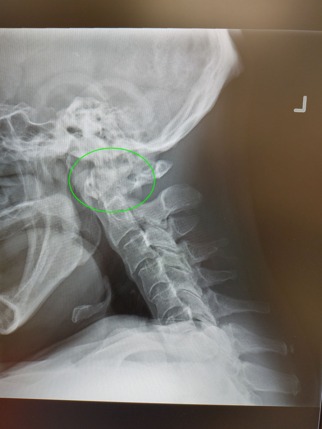
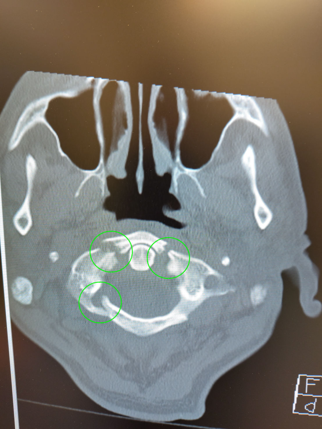
       - C1 – appears cloudy. Radiologist report – no findings 
       - No changes in pain 
       - Oral hydromorphone 1mg given

Admitted for pain control and head CT 
C1 – Jefferson fracture 
       - Canceled admission to hospital and rerouted to specialist care. Failure to improve over time with different management 

Learning points
       - What does a vertebral fracture vs soft tissue injury look like clinically
              o Neurological 
              o Rigidity and persistent after treatment
              o Disproportionate pain 
              o Concerning mechanism 
              o Concerning patient features - Age, History previous fracture, Predisposing conditions making fractures more likely
       - How urgent is imaging in this patient
              o Not overly urgent – trying to detect people who are at risk for secondary injury after initial injury. 
              o Minimize movement 
       - What is the most effective way of immobilizing suspected spinal injury
              o Listen to what the patient is telling you. Don’t apply anything or move them if they are telling you not to. 
              o No strong evidence in using c-collars as being very effective to immobilize.
              o Have person inform you of what their optimal position of comfort is. 
              o If unconscious use head log and place in somewhat anatomical alignment. 


Gallery


CASE 1
Fly flew into females ear and she can still feel it. Tympanic membrane – clear. Could see fruit fly in ear canal
       - Lay patient down with affected side up to make a cup and fill with lidocaine for a few minutes.
              o Anesthetized ear a bit
              o Neurotoxic to bugs
Flip patient over and drain ear and see what comes out. Nothing came out – used elephant ear washer and flushed out bug.

CASE 2
Male mowing lawn and feels sharp pain in forearm and sees a spider jump off. Came in next day with 3cm diameter bruising and swelling to area.
       - Ultrasound – nodule under the skin
Unless spider is venomous – most care is supportive.
Questions to consider
       - Is the spider currently residing in Australia or imported into another country
              o Watch and consult quickly with toxicology for anti-venom
                     - Funnel web and redback spiders dangerous
       - Is the spider native to North America?
              o Brown recluse
              o Black widow – black body and red hour glass on body

☙ Brown Recluse (AKA Fiddle backs)
       - Like to hide
       - Live in shadows
       - Bite has hemolytic effects and localized necrosis
       - No anti-venom
       - Treatment is supportive
       - Treat for secondary infection
       - Cold compress – slow down toxin – don’t give frostbite
       - Don’t typically live in Canada
☙ Black Widow Spiders
       - Envenomation that can be dangerous
       - Variable presentation
       - Treatment is based on speculation and anecdotally
       - Anti-venom available in Canada
Patient – localized infection. Pt to monitor if wound got worse.

Rural Surgical
Case 3
Pediatric appendicitis
       - ALVARADO score, WBC, history, and exam is difficult to predict.
8 hours RLQ pain, walking with shuffle and mom brought into ED. No fever or elevated WBC
Ultrasound – found cecum, pouch measuring 6mm in diameter
       - Consult with Surgeon – and went to OR for appendectomy.
Sent home next morning.

Case 4
Using table saw and cut of tips of two of fingers. Index finger sheered off 2mm distal phalanx – worry about osteomyelitis but phalanges are exception to rule. Get out Rongeur to nibble away at phalanx and close with skin.
       - Did not completely cover with skin but enough to have subcutaneous tissue cover and skin will heal over with secondary healing. Maintain the length of digit and nail bed and preserve function.

Case 5
Roofer who shot nail from nail gun through boot into foot. Hard to determine where the nail is due to steel toe boot.
- Sedated to remove nail from boot and foot.
- Went through soft tissue – no bone involvement
- Monitor for infection


Nausea, vomiting, diarrhea

☙ Top 10 presenting complaints
       - Largely due to poor coping skills at home
       - Rarely due to severe condition.

☙ Vomiting without red flags (listed below)
       - Unstable or septic
       - BGL
       - Hematemesis
       - Fever in returning traveller
       - Community outbreak
       - Chronic – greater than one week
       - Up to 3 months old

☙ Stable patient
       - Stable vitals
       - Drinking – whether able to keep fluids down
       - Looking for IV fluids
       - Can be rude
IM dose of antiemetic – first line is ondansetron 4mg IM
       - NOT IV
       - No blood work or IV fluids

Children – no diarrhea – gravol (dimenhydrinate) suppository. If diarrhea then IM ondansetron
IM – metoclopramide

Set 30 min timer

☙ QT prolongation – don’t usually worry about it unless known cardiac history on cardiac meds. Never give 8mg at a time and if requiring more in 8 hours its usually divided into 4mg/dose. Rarely need more than 4mg.
       - Review previous ECG to look at QTC. If none available order ECG to review.
       - Pediatric dose - 0.1mg/kg up to adult dose of 4mg.


Evaluate patient after 30min
       - History
       - Physical exam
              o Expected to be benign
              o Diffuse generalized abdominal pain
       - Pinpoint severe peritonitic pain
              o Changes workup around umbilicus, RLQ, gallbladder, pancreas
              o Abdominal ultrasound to rule out any areas of concern

Vomiting status one of two situations
1. Vomiting resolved
2. More through exam and history and rule out any red flag concerns listed above. additional 4mg ondansetron if still not settled vs giving other antiemetic such as gravol.

☙ If ongoing vomiting for days and refractory to dimenhydrinate or history of cannabis consider
       - Dex 4mg by any route effective 45min – 6hours.
       - Haloperidol – 2.5mg – cannabinoid reaction
Consider blood work
       - CBC, electrolytes, creatinine,
       - VBG, lactate, extended electrolytes and TSH in frail and elderly
Look to cause of vomiting.
       - Usually viral infection
       - Food poisoning
       - Drug history
       - Exposure to poison
       - Other acute condition

☙ Most patients will feel better after 1st dose and second dose.
☙ Wait 30 min post vomit to take medications with small sip of water. When vomiting stomach loses its ability to tolerate large amounts of fluid and you need to take small sips of water (5ml) every 5 min over an hour and then increase to 10ml every 5 min. If vomit again start over.
☙ Use other fluids – coffee, milk, juice, Gatorade,
       - Pedialyte no better than other fluids listed.

Educate on home management
       - Go by over the counter gravol (consider suppositories – unless having diarrhea)
       - If tried and failed to manage at home – prescription for ondansetron.
☙ Not a big fan of normal saline due to pH being 5.5. Lactated ringers pH 6.5
☙ Patients better to replace fluids through their own GI tract than IV.

Moderate to severe symptoms – requires different management.
       - Blood work – unless underlying condition or severity of presentation – don’t usually order.

☙ Vomiting patients absent of red flags – rarely have any abnormal bloodwork
Benefits of vomiting
1. Difficult pt with unmanageable symptoms and get them feeling better without tying up the physician and allows for more smooth interaction
2. Invest time educating low risk patient
3. Avoid over medicalizing
4. Send pt home feeling better and better prepared to manage vomiting in themselves, family, or friend in community.


Learning Objectives

☙ Approach to seizing patient 
☙ Managing DKA
☙ Arranging Transport 

Case
EMS transport – no pre-alert came in ventilating patient
       - Witness seizure approx. 1min
       - Took photos of medications - - Ramipril, allopurinol, Jardiance, nifedipine 
Call in additional support – General Practitioner Anesthesiologist (GPA), emerge RN and two LPNs and had to call in lab and x-ray
Defibrillator Pads place and IV's started

Airway - as per EMS - NPA difficulty bagging as patient has large neck with vomit everywhere with SPO2 in upper 80s
Breathing – looks deep and rapid (kussmauls) course and decreased to bases bilaterally
Circulation – hypertensive 195/110 p 130s warm extremities and afebrile
GCS 3 – pupils 2mm minimally reactive
BGL – unreadably high 
No obvious injury 

General seizure starts
       - Ativan 4mg IV followed by another 4mg dose given 4 min later
       - Phenytoin 20mg/kg – seizure stop
Total seizure 5 min. Patient hypoxia – blue in color difficult to ventilate

LABS - VBG, glucose, electrolytes, osmolality, LFT, CBC, tox screen, CK, creatinine, and lactate 
ECG – narrow sinus tach and QTC 570 no ST-elevation
3 total IVs placed

Family arrives but provides minimal information other than states patient doesn’t drink much alcohol

☙ Priority is to manage Airway 
       - Fluids
       - Vasopressors – norepinephrine 
       - Equipment 
☙ Consider the challenge of staff and their scope of practice from RN to LPN and try not to overtask anyone

GPA – intubate – no paralytics or pre- procedural sedation 
       - Started propofol post intubation 

Blood gas – pH6.8, bicarb 8, lactate >20 – No PaCO2 available

Consult with transport Doc – who is also helping with pt management 

Vitals Pulse 130s, BP 130/80, post intubation SPO2 92% on ventilator PEEP at 5 with increased RESP rate to blow off acid. 

Secondary survey – unremarkable except continue course bilateral breath sounds. 

BP decreases 80/50s 
       - Pressure bag of normal saline 
       - Pressors - norepi - 0.1mg/kg/h

Setting up ART line and central line being set up

More labs 
WBC – 20 
Hemoglobin – normal
Na – 133
K – 3.9
Anion 39
Creatinine 159
Glucose 51
Lactate 25
LFT normal
Serum osmolality – 365 (results received next day)
Chest x-ray - no consolidation 

Start DKA protocol – most centres have printed form.
       - Monitor potassium and supplement
       - Monitor fluid resuscitation
       - Insulin infusion - 0.1Unit/kg/h
       - Monitor electrolytes and anion gap regularly.
       - Cover infection – Piperacillin/Tazobactam
       - Consider treating alcohol poisoning – Fomepizole

Limited number of people to admin med and IV routes. Need to prioritize order.

Another Seizure 
       - 4mg IV Ativan
       - Keppra Loading dose drawn up 

Repeat Blood gas – pH 7.17, bicarb 13, and lactate 15. BGL still unreadably high

ART and central line in
Vitals stable. Improvement with metabolic acidosis. 

Transport considerations 
- Two provinces on the call 
- No air helicopter
- No flights 
- Only ground transport
- Decision was to send ground ambulance with GPA to meet with specialized team.

Repeat Blood gas – pH 7.22, Bicarb – 17, Lactate – 10

Chest x-ray – confirm central line, Foley catheter, Check all IVs to ensure patency. 

5 days in ICU. 8 days in medical ward
LP, EEG, CT Head – normal. Thing seizures were from severe DKA. Did end up with aspiration pneumonia. 
Treated for Type 2 diabetes – A1c - 16. Was previously being treated months prior but stopped because he didn’t want to take meds anymore. 

Resources 
- EM cases podcast and summaries used to help with resuscitation
- Resuscitation crisis manual

Reflection/challenges
       - Nursing allocation of task 
              o Close loop communication
              o Don’t overwhelm with multiple orders at a time
       - Prioritizing tasks 
              o Limited people and interventions
              o Would have wanted more fluid resuscitation, got sidetracked with seizures/other medical problems. 
       - Communicating between team and specialist on phone
              o Multiple people on call
              o Spotty services
       - Multitasking
              o No time or space to reflect on what is happening
       - Deciding when and how to transport the pt
              o Hard without air support



 Simulated Rural resuscitation practice – there are multiple courses offered throughout Canada 
☙ ATLS, PALS, ACLS - need to review for high acuity calls in limited resource settings constantly
☙ Practice regular rural resuscitation in person and virtually to help keep skills up          

Air transport

Takeaways:
☙ Remain objective
☙ Risk associated with transport medicine when untrained.

Advocate for optimal patient care—Sometimes, this can be the opposite of advice given by a specialist.
       - Have open and constructive communication.
       - Question if there are dose differences to make sure there isn’t a communication error

Generalist opinions are not inferior to specialist opinions—they are collaborative, so raise concerns if something doesn’t fit.
“The first one to plead his cause is right until his neighbour examines him.”

CASE
20yo with TBI moved to tertiary centre 900km (2.5h flight away)

☙ First consult – neurosurgeon requests patient not intubated and keep that way if possible. ---Stated that intubation will increase ICP.

I picked up the patient and explained to the ED physician about the plan – the patient was on propofol and midazolam. Pt fought sedation the whole time.
       - The flight was through a thunderstorm. Unable to monitor pt properly.
       - Intubation at this point is impossible due to turbulence
       - Ran out of propofol – had to change medications.

The nurse and RT at the receiving hospital questioned why the patient was not intubated and was preparing to intubate before leaving.

With gaining experience over the years would have questioned the orders better.

**Understand your knowledge gaps and find ways to improve education.
☙ Some provinces do not have transport services that support patients. Sometimes, you might think you should go on the transport, but use caution.
☙ If you go on a transport, you are entering an unknown environment, have no idea where any supplies are, and are not working with the typical team you are used to. This causes a safety hazard to the patient with not fully understanding the environment.
       - Best to wait and stay in your environment and wait for the proper transport team to arrive.


Case

EMS brought in 62yo male post-arrest hooked up to monitors in the ED. Sudden collapse found by a bystander and 7min EMS response – worked patient for 10 min for an estimated total downtime of 17 min.
BMI 21 weight - 60kg
No significant med history
Acting normally before collapse.

Vitals
☙ BP - 110/90
☙ Pulse - 92
☙ Temp - 36.4
☙ Igel – resp BVM -12
☙ SPO2 -100 on 10l
☙ ETCO – 25 good waveform

Pupils fixed and dilated @ 10mm with no reaction
Agonal resp no purposeful movement
Consult to move patient to tertiary hospital – receiving physician asking for intubation for transport. 

Anatomical airway checks – good

Induction medication
       - Ketamine (reduced dose) 1mg/kg – 60mg
       - Rocuronium – no concerns with prolonged paralyzation (60mg)
       - Succinocholine – unknown what potassium and renal function is – not used.

Not a time-sensitive urgent intubation. No anatomic or physiologic contraindications
       - Patient pre oxygenated
       - Position on the stretcher is good
A – Direct look
B – video look
C – fallback to supraglottic airway
D – cricothyrotomy

☙ Post intubation, Ketamine and Rocuronium will provide a 30-40-minute window to start infusion or prep ongoing boluses.
☙ If a ventilator is present its good to have it on standby if transport is not readily available

Have airway equipment checklist – use your own that you are familiar with and have *modified based on experience.
Equipment:
       - Suction
       - BVM
       - Ventilator
       - Ongoing sedation plan
       - O2 source
       - Syringe for inflating ET tube
       - Lyrigoscope – proper blade and functioning light
       - Backup airway – iGel, oral airways
       - ET tube holder
       - ET tube

NIBP – now dropped 62/30
Pulse check – unknown
EMS is still on scene

POCUS – to confirm cardiac functioning. Clearly beating. LV beating well and squeezing well. Good stroke volume. Mitral valve, not a full excursion. EPSS – Ejection fraction around 40%
       - Don’t start CPR in this case it will hinder pt care
       - BP likely due to EF of 40%

Intubation is no longer a priority at this point – Cardiac stabilization is now the priority. 
- Need to improve Mean Arterial Pressure (MAP) to neuroprotective levels 

MAP target – min of 65 for adults at risk to protect the heart. Min MAP of 70 to protect the brain.
MAP of 70 target for this patient. 

Neuro –In closed head injury, cerebral perfusion pressure (CPP) equals MAP - ICP. normal ICP is 7 -15 mmHg. This pt could have a head injury (with blown and fixed pupils bilaterally) with increased ICP. If the patient had ICP of 40 – 70-40=30

Current MAP 41 – use POCUS to rule out inferior vena cava – no collapsing completely – no preload dependent. IVC is not fully distended – not obstructive shock
- No clinical signs of bleeding or fluid loss – FAST scan

Consider cardiogenic shock (known element in this case) and neurogenic shock
Neurogenic shock – depressed brain stem - dilated pupils – consider vaso pelagic and blood pooling in legs and not returning to heart as well as could be. 

Do you want crystalloid? (pt received only 200ml Lactated ringers at this point)
- No signs of overload (can use ultrasound to check lungs for pulmonary edema) reasonable for 1L bolus of LR and reassess
- Cardiogenic shock management – assists the heart in returning to normal parameters of function. HR and BP within normal ranges. Target into the normal range

Think about heart function in 4 steps.
1. Preload – want preload optimized – meaning plenty of fluid upstream for heart
2. Chronotropy – how quick is the heart beating optimal 60-80bpm
3. Inotropy – how much is heart squeezing (Frank Stirling) – can use vasopressor
4. Afterload – MAP of 70 – don’t want excessive as heart is having to work harder to overcome resistance. 

Vasopressors 
       - Norepi – good choice - more alpha effects improve preload. Also has beta effects, which increases inotropy through actin myosin troponin system. Good for supporting cardiac activity. Does take time to mix and get running.
       - Phenylephrine – pure alpha 1 agonist. Help with preload. 200mcg bolus (large dose) 
       - Repeat BP – Map improved 41 – 62

If critical BP, start with a larger dose. If BP/MAP ok then start with lower dose (norepi starting dose started at .05mcg/kg/min). The MAP doesn’t improve with the initial nor-epi dose and after 3 min MAP still at 62 – increased norepi dose by doubling the dose to 0.1mcg/kg.min and MAP increase to 67 - let BP recycle over the next few min and MAP decrease to 64. Double nor-epi dose again to  0.2mcg/kg/min – MAP improved and maintained at this dose. 

Intubation now that BP stabilized 
       - Ketamine – SIVP that can sometimes destabilize cardiac function
       - Rocuronium 

Tube confirmation
       - Tube misting 
       - Auscultate bilateral apex
       - ETCO2 
       - Secure tube

Sent Janelle went with EMS and premade syringes of ketamine 50mg/ml – 1ml q 5 min if needed and phenylephrine 100mcg/ml q 5 while titrating nor-epi. 

Try to catch changes early and manage with smaller doses versus having big swings in patient stability and struggling to catch up


Discussion of what it is like being a rural physician with limited resources.

Objectives:
- Challenges in low-resource settings
- What is a resource-setting
- Lessons learned working in low-resource settings
- Review and compare the management of cases in resource-abundant and limited settings
- Discuss different cases
- ED peer-to-peer programs

What is a low-resource setting?

- It means different things based on where you are. One site might have different options compared with another site.
- There can be a lack of staff, high turnover, locums coming and going, challenges to continuity of care, and a lack of equipment, imaging, medications, specialists, and experienced practitioners. This is not just for remote areas. Consider urgent care centers in Calgary, AB – run by GPs with some imaging capabilities (x-ray), limited lab, no specialists, but still considered an emergency department.
- Rural vs. Remote: Rural areas refer to areas with a lower population and less infrastructure and might be more accessible with clear routes to urban sites. Remote areas are distant or hard to reach due to geographical challenges with less clear routes to urban sites.

Challenges working in low-resource settings

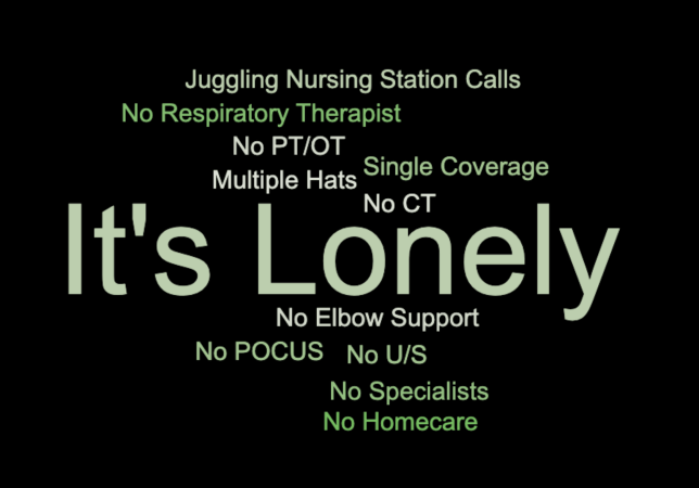
- The main problem is that it is lonely, with no colleagues nearby to lean on for support -No elbow support.
- No PT or OT, no RT. Your managing intubation and supporting pt aftwars. Huge reliance/dependence on transport and at the mercy of weather as well as transport crew schedules (pilot timeout). No imaging support.
- Wearing multiple hats – ED doc to hospitalist if you are the only physician on and need to admit a patient. Learn to test wisely vs sending them out – consider admitting and observing over night vs transport.

Lessons Learned while living in low resource settings

- Learn to live with uncertainty but realize that help is only a phone call away
- POCUS skills are paramount.
- Rules of care and standard of care change based on location.
- Changing practice and location – helps with burnout.
- Know where you are transporting to. Depending on where you are in remote settings closest ICU may be hours away and require multiple modes of transport.
- Might have to use a community pager – where other allied health care call looking to consult for support.

Review and compare management

- Tylenol OD with no labs – draw the blood and send the patient out to a level 1 facility. *start the treatment* you are not going to be able to wait for the labs to come back because they have to be flown out – likely with the patient.
- Diverticulitis or renal colic with no imaging without an acute abdomen. Bring the patient back the next day to reassess. If the patient's condition worsens, then transport.
- Appendicitis – used POCUS – sent photo to the surgeon to consult and recommended patient transported.
- Compartment syndrome – fasciotomy with ortho consult and permission on the phone and through video.
- Airway management – learn ventilator and know settings. You will miss RTs
- Sepsis – start antibiotics, blood work and cultures – come back in a couple of days.
- Form 1 – Consult psychiatry to determine if the form can be lifted or maintained.

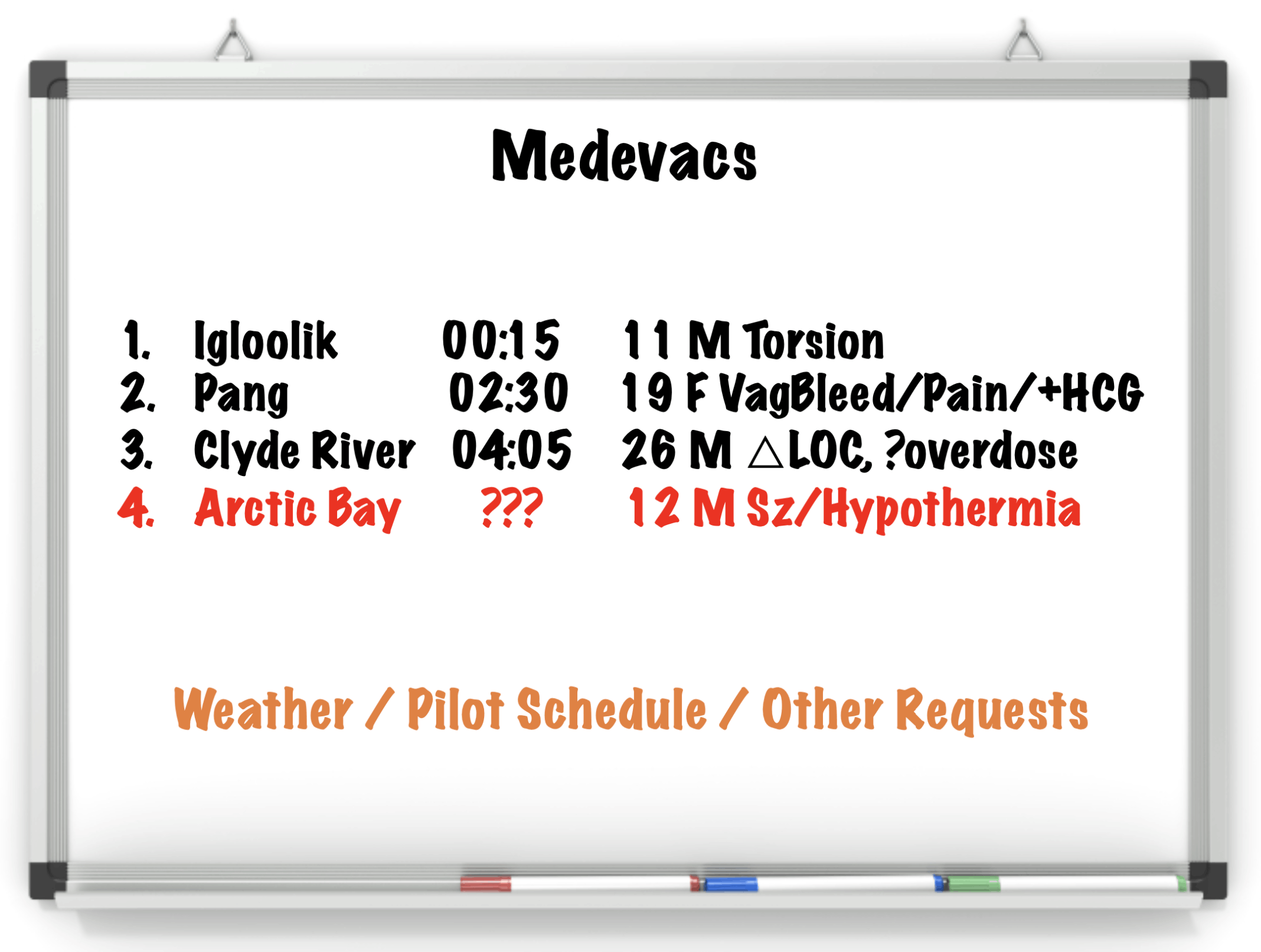
Discuss different cases (see photo below)

- Medivac vs schedivac
       o Medivac needs to happen ASAP.
       o Schedivac – scheduled appointment.
- prioritize patients transports and bypass to more definitive care if needed. 

ED Peer-to-Peer
- Call another ED colleague to receive collaborative support.
- The challenge is that rural practice is lonely. Recognized the need for 24/7 support and that more practitioners might be willing to take rural shifts.
- Two heads are better than one, and it is nice to have someone not directly involved in the case providing feedback.
- Helped to keep people in the community and avoid unnecessary travel.
- Consults can vary in complexity and sometimes can be just a simple review of a case.
- There is an increase in the uptake of locums, and practitioners report feeling more supported in the community.


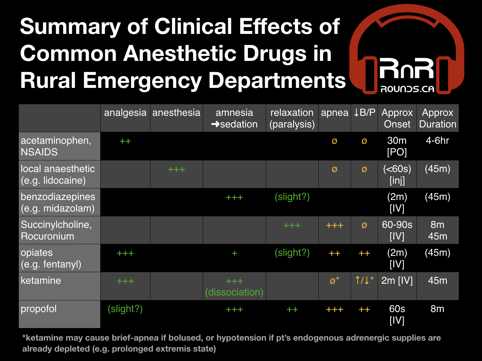
RxAnestheticTable

This table is provided as a sample based on personal experience. 
Exact numbers and properties will vary depending on the source.

EMS brought in a 62-year-old female with altered LOC from a motel room surrounded by pill bottles, blister packs, and a bottle of helium. It is thought to have been ingested within the last two hours. The patient had previously admission to the hospital for suicidal ideation. EMS and police brought all medications, and the staff was able to identify 10 of them.
 ☙Perindopril, primidone, sertraline, rosuvastatin, trazadone, b12 quetiapine, aspirin, bisoprolol, and Ativan.

Bisoprolol - most concerning.
☙ HR 70
☙ BP 110/65
☙ R – 16-18
☙ Sp02 – normal – no supplemental O2 given at this time.
☙ T – normal
☙ BGL – normal

Patient initially protecting airway, answering 1-2 words, but increasingly became drowsy.
☙ ECG, IV, Fluids

Estimate tablets were taken and consult with poison control.

HR and BP did start to decrease. Lactated Ringers bolus given vasopressors drawn up. Intubation due to increasing drowsiness, and vitals trending wrong way and suicide note left and intentional overdose. Narcan was given to rule out possible narcotics.

Poison Control – Priority treatment for bisoprolol (21 tablets at 105mg each estimated to be ingested) and concern for hemodynamic consequences.
- Suggested decontamination with charcoal via NG tube due to believing pt ingestion was around two hours before arrival. 125g activated charcoal to start.
- Bolus of IV fluid, and if BP continued to decrease, to give norepinephrine.
- Calcium Gluconate – two amps.
- If Norepinephrine is ineffective - start with high-dose insulin therapy – giving a bolus of 1unit/kg of regular insulin and following up with a continuous infusion of 1unit/kg/hour if no improvement in hemodynamic stability titrate up a unit/kg/hour, every 20-30min to a max of 10units/kg/hour. Given with dextrose as well (rare to see glucose drop precipitously – monitor glucose and serum potassium)
Insulin used to treat heart metabolic problems not glucose in this case – has positive inotropic effects and stimulates myocardial glucose metabolism and lactate intake
- Primidone, trazadone, Ativan – sedatives – concerns for respiratory status – managed with intubation.
- Aspirin – monitor for toxicity – in this case, initial and repeat serum aspirin levels were negligible.
The patient was not on high-dose insulin for long – DC in ICU.


Initial management of patient considered in steps:
1. IV, O2, monitor, glucose, ECG, IV fluids
2. Decontamination with charcoal
3. Starting Vasopressors
4. High dose insulin
5. Lipid emulsion therapy – did not get to steps 5 & 6
6. ECMO

Learning Points
1. Many drugs can produce low and slow toxidrome (bradycardia and hypotension) – Beta-blockers, calcium channel blockers, digoxin, opioids, clonidine, and drugs that fall into the sodium channel blockers category. If seeing a patient with bradycardia and hypotension without concerns of toxin consider – hypothyroid, hypothermia, post MI, spinal injury, hyperkalemia.
2. Activated charcoal is advised with beta-blocker OD if it is within 1-2 hours of ingestion.
3. Specific treatment - Calcium is recommended for cardiac membrane stabilization and high-dose insulin. Insulin bolus is given in tandem with D50W then 1 unit/kg/hour. Continue to monitor glucose and potassium.
4. Vasopressors may be indicated. If you can do POCUS and notice decreased heart contractility, consider epinephrine. If contractility is normal, consider norepinephrine.
5. Lipid emulsion therapy – on poison control website
6. ECMO – last step consider transport options early if not located close to site with capabilities


CASE:
30yo female brought in by EMS seizing. 39 weeks pregnant – Eclamptic seizure till proven otherwise.
The roommate heard a thump and witnessed the patient size – approx. 30s tonic activity, no clonic activity. 

EMS – semi combative – 5mg midazolam given. The patient vomited – 4mg ondansetron was given. 

ED – partly sedated and partially agitated. 
Unable to obtain NIBP due to patient agitation
P - 123
SPO2 -98%

Magnesium 2g over 5 min using syringe due to poor IV site. While attempting other access managed to obtain 4 more sites. 

CBC, lytes, creatine, LFT, INR
Consult obstetrician – LDH, urate, fibrinogen, haptoglobin
2g magnesium was administered again

Total 4g mag 

Still agitated and requiring restraint. 

30mg ketamine given – with minor effect
20mg ketamine given – Patient settled down – NIBP 125/95

Put in foley catheter 
Obtained fetal HR – 160
NST machine brought down – good baseline with slightly decreased variability, which is not unexpected with giving the magnesium.

Ankle clonus when compressing ankle 6-12 beats
Patient starts to come around and is more responsive – confirms no prenatal care sought out.
Patient not found in provincial records. Family physician at site was helping and able to find some records of her coming with concerns of being pregnant (which were positive) and no further follow up of any further care found.
24 hours later, patient delivered.


Medscape review
☙ The main priority is to stop seizures
       o First-line drug - Magnesium 2-4g given over 2-5 min
       o Benzos (midazolam or lorazepam) 2-5mg IV over 2-5 min
       o Phenytoin – if mag is contraindicated (hypermagnesemia), benzos and phenytoin become the main drugs
☙ Second priority control hypertension
       o Systolic over 160 or diastolic over 110
       o Hydralazine 5-10mg over 2 min or labetalol IV 20mg
       o Goal is to maintain BP systolic 140-160 and diastolic 90-110
☙ Supportive care
       o Consider the need for intubation – especially if given lots of benzos
☙ Definitive care for pre-eclampsia or eclampsia – is Delivery if not managed medically
☙ Consider steroids for preterm deliveries, BUT consult before giving as there is a narrow window to deliver the baby after the medication has been administered.
***Consult with the obstetrician***
☙ Do not want to give more than one dose of steroids, and delivery should happen within 24 hours of steroids.
  - Small window of opportunity to delivery
      o Betamethasone 12mg IM q24 hours x 2 doses – ideal
      o Dexamethasone 6mg IM q12 hours x 4 doses
☙ Premature rupture of membranes – Give steroids. Still, consult with OBGYN


Laura Dunkin

Two patients arrived both approx. 20yo. One male on female by EMS
Decreased LOC. Respirations ok. No obstruction. Unknown how long they were presenting like this. Mom found them sleeping in the basement and couldn’t be roused
Soft BP around 100 systolic and lower HR in the 50s

Priority is to triage 

Female resp rate decreased 8-9
Male resp 12-14 

Narcan by EMS – no effect
BGL – normal
Pupils normal
GCS 7 – female.
Afebrile

Repeat Narcan – no response
☙ IVs established and some fluids given due to soft BPs
☙ Labs – VBG, septic workup, serum and urine 

Male – more stable GCS, not as severe, with good respirations.

Female intubated – set up transport. 
☙ Intubated with roc and ketamine – airway clear 
No indications for narcotics or alcohol use

Friend talked to pt mom and informed them they took GHB and mom called hospital. Transport in timely manor

Outcome for female – extubated later that night and had a good outcome
   
GHB – used to increase intoxication feeling.
- Less calories. Common in bodybuilders to enhance intoxication without additional calories of alcohol.

Patient - young man brought in by EMS CPR in progress (during COVID). PEA on monitor – no vitals. Appeared pale and cyanotic – EMS had a suicide note and a bottle of sodium nitrate from the patient's residence.

Staff were able to contact a colleague who was a toxicologist specialist for advice. It took an extended period to get poison control on the phone. The patient was intubated, and multiple lines started.

Recommended starting Methylene blue started (color of blue ink) started running right away and continued with CPR. The patient started turning blue as infusion began circulating. Redosed q 5 min (5-6 doses total) – after 45 min of CPR patient remained in PEA and time of death was called.

Concerns for how the patient was able to obtain it and suspect possible online purchase? Unknown how patient was able to obtain sodium nitrate

☙ Sodium Nitrate (nitrates) causes methemoglobinemia. The Fe2+ (healthy) atom in hemoglobin is oxidized to Fe3+ rendering the molecule unable to carry oxygen efficiently. Methemoglobinemia turns RBCs a chocolatey brown colour but seen through the skin, appears as a profound cyanosis
☙ Methylene Blue treats methemoglobinemia by reducing the Fe ion back to 2+. Thus despite being a richly blue colour itself, it actually helps to resolve cyanosis in this context.


080 - How to become a better Rural Resuscitationist  

What is Resuscitation – making clinical decisions based on limited time and information – gambling essentially.

The sicker and more unstable they become, the greater their risk.
Be comfortable with making important decisions with limited information and accepting the risks you may make the wrong decision and that sometimes there is harmful outcomes
       - Know that failure to act and decide is also a choice and has its own consequences.

Be aggressive when resuscitating – especially pre-arrest patients
       - Delay in treatment and loss of pulse results in damage control and trying to recover with minimal brain damage if possible.
       - There are no black-and-white recommendations in pre-arrest patients
       - there is a risk of having underlying causes that lead to death based on the treatment given.
       - Identifying anything time-sensitive - don’t delay
Ex:
☙ Anaphylaxis or sepsis – once identified or high suspicion – treat!
☙ Toxicology – treat with activated charcoal if possible
☙ Toxic shock – remove (replace if needed with clean dressing), use caution with abx in this case.
☙ Critical Bradycardia – give atropine.
☙ Critical hypotension – start a vasopressor (whatever you have or are comfortable with to start) to stabilize and switch to a different pressor afterwards if needed
☙ Look at H’s and T’s for initial diagnosis and treat quickly in ACLS

Slower decline patients in delirium
       - Small dose of ketamine to help sedate 
Phoning a friend – causes delays of min 5 and greater – attribute the harm that comes with delays.

Protocols of resuscitation
IV fluid – 2 L of fluid – much time spent on what fluid to give and how fast to run it in – Don’t delay other treatments on fluid. Other times, there are other more effective treatments than just fluid.

☙ Think ahead to be ahead – while waiting for transport, think about what is coming next – transport. Try to be two steps ahead of where the patient is at. What might the patient need next – set up for airway, set up for vasopressors and pump programmed ready to go, get d-fib pads on if cardiac issues arise (ex. STEMI or arrhythmia patients)

☙ Think about barriers to transport!
       - What can you do to expedite the process? Team arrival is not the saviour.
       - It’s the destination the patient is going to.
       - The goal is to help the transport team as much as possible.
       - Delaying basic interventions can be detrimental to patient care and delay transport to definitive care.
Ex. What drugs are the patient going to need and get them going prior to transport? If you can give them meds before transport and reduce the amount needed in transport, that would be better.
       - If IV is not reliable, then start another. Consider a central line or IO.
       - Heavier sedation during transport is normal due to the amount of stimulation experienced during transport.

Resuscitation is rarely black and white where you know 100% what to do. Get comfortable with having a limited picture and play the odds and probability of the patient's best interests.

☙ Analysis Paralysis is how you lose at the resuscitation game.
☙ Get comfortable with being uncomfortable


Case

55yo male previously diagnosed with major depressive disorder (MDD) being treated with fluoxetine and going through marital problems with worsening mood.
       - Took 10 -15 tablets immediate release Asprin 325mg – no co-ingestion

Time to assessment is 2 hours post-ingestion and feeling at baseline during the exam. Vitals normal. Physical exam normal. No activated charcoal was given due to the time elapsed.

☙ Toxic ASA levels 150mg/kg – pt toxic dose is 12,000mg. Pts reported dose is 3000mg based on the 10-15 tablets reported.

Patients Serum level - 3.65mmol/L reported back from the lab – Critical value.

☙ Management
       - If no toxicological level is available but the patient is presenting with s/s of:
              - OD (ex., tinnitus, hyperventilation, vomiting, seizures, respiratory distress, hyperthermia), these are good clinical clues.
       - ASA level of greater than 3.5mmol/L
       - Presence of metabolic acidosis

***Endpoints of therapy (3 points that all have to be met)***
       - ASA level of less than 2.2mmol/L
       - Two consecutive ASA levels of declining trend
       - Clinically patient must be doing well

ASA OD – thinking ahead
       - If you think it is serious – start arranging for dialysis ASAP
Call Poison Control – alkalinization process of urine
       - Prepare bicarb drip – take 1L D5W and remove 150ml and use 3 amps of 50ml bicarb to replace fluid then run at 1.5 times fluid maintenance rate up to a max of 200ml/h.
       - Monitor urine output of 2-3 ml/kg/H – TARGET output
       - Insert a foley – empty urine in the bladder for a baseline for patient's physiology and monitor exact volume and urine testing.
              o Check urine q2h for pH and serum blood gas, potassium, and salicylate levels.

Specific targets are harder to get the more serious the cases (which is why dialysis consideration is so important)

☙ Targets
 Urine pH >7.5
 Serum Potassium < 3.5-5 role is for the urine alkalinization process.
 Serum pH <7.56
 Serum salicylate aims for <2.2 with two continuously declining levels

At the 5–6-hour mark, the repeated serum level was 3.92, and the patient started getting nauseous, diaphoretic, tachypnea, and tinnitus, and resp alkalosis (at worst presentation)
Within 36-48 hours, the patient was treated effectively and resolved all levels. No dialysis was required.


Buffalo Attack

It is a very remote site, a 5-hour drive to tertiary care. Patient is two hours further away from the site.

Buffalo throws the patient to the ground, and the patient lands on his back, the buffalo hits him in the chest with his head, then gores the patient to the left chest (around the 6th rib midclavicular). The patient also gets gored in the back of their legs.
       - Large open chest around 20cm x 10cm following along the rib with lots of red subcutaneous tissue exposed (no sucking chest wound). Concerned for femur fracture in the left leg.

Pt has minimal pain with minimal analgesic (100mcg fentanyl over 2 hours). Spo2 95% HR and BP with in normal limits – no signs of early shock

50yo male with a higher BMI of around 33 and otherwise healthy.
       - ABCs normal. No head or neck pain. Only pain to central chest (not the gaping chest wound) and back pain and pain to the legs

Priorities – transport to greater care.
       - Concern for pneumothorax – ultrasound had no lung slide but doesn’t necessarily mean pneumothorax when you can’t see the two plural lines moving opposite each other.
       - Ultrasound is sensitive to pneumothorax - look for lung point – on ultrasound you can see one side apposition of plura and see it sliding and the other side where you see the absence of sliding. Looking where the visceral pleura is peeling off the parietal pleura. Tracing out the margins of that, you can see how large the air bubble is.
       - He does have a small pneumo – so consider treatment – does patient really need large chest tube? Used small pigtail in this case.

Central chest pain – ultrasound used to look at bony context – found sternal fracture.
Concern for internal bleeding – placed patient in Trendelenburg to see if there was any fluid in the abdomen. It would be caught in the area of the diaphragm to see early detection – nothing major found.
Long bone fracture – linear probe used to follow the cortex of the bone. Not superior to X-rays in long bones. No fracture was noted.
Heart – able to look in subxiphoid and parasternal – no pericardial effusion or mechanical issue. No signs of obstructive or hypovolemic shock.
Within a short period of time, ultrasound was able to rule out very dangerous concerns
CT scan available – verified all found on ultrasound.

Take home points
1. Stay away from wild animals! Fatalities typically come from species that don’t get the attention. It’s typically from the “cute and fuzzy” animals.
2. Consider the amount of force that these animals can cause.
3. ABCs, ATLS and getting pt set up for transport most important.


RxAnestheticTable

.

CASE - 28yo female brought in via EMS with GCS 3 who was at a bar with friends and family, she went outside to have a smoke and was found unresponsive (unwitnessed) in the middle of the afternoon. No suspected foul play.

No meds or med hx
Vitals with EMS
P – 88, Spot 98% on RA, T – 36.8, BP – 120/80, R – 16

No known risk factors – no drug use, was out with friends and family had some alcohol.
Well dressed, well groomed, no track marks

Exam: No muscle tone, pupils slightly miotic (but not obvious) but reactive. Full breath sounds, no snoring resp or vomitus. Strong pulses, chest auscultation unremarkable. Abdo is soft, the neck is supple, no obvious trauma, and moist mucous membranes. No spinal concern – no step-off concerns. Good rectal tone. No discernible reflexes 

Appears to be in a deep sleep.

Review ABC’s, IV access, airway stable at present.
BGL – normal, empiric Naloxone – started with .4mg and increased to 1mg up to 2mg no response. 
CT scan – normal - Patient intubated after scan 
Labs – VBG lactate, extended electrolytes – all normal. Tox screen came back after patient intubated with Ethanol  118mmol/L

Consulted with ICU – review of common issues - hypoglycemia, intracranial issues, opioid overdose? None found.

Collateral history - Sister reported to EMS that patient had only been drinking alcohol with no other substances. Reports that patient is a heavy drinker (family unaware)

Sedated with low doses of propofol and fentanyl – patient started waking up quickly and decided to increase sedation while figuring out story. Patient later extubated in ICU once no other concerns cleared and story continued to check out.

Toxic Alcoholism typical concerns:
- Tachypnea – underlying metabolic acidosis – till VBG comes back
- Seizures, especially if serum tox can confirm.
- Serum tox screen ethanol and serum osmolality.
- If no CT - need transport to rule out.


076 - Serotonin Syndrome - Tox 2 miniseries

Case - 32yo male with a history of Major Depressive Disorder(MDD) and taking Sertraline and bupropion.
       - Referred to ED from PCP presenting with loose diarrhea for last 10-12 days – concern for electrolyte abnormalities
       - No complaints of fever, vomiting, abdominal pain, appetite intact, jaundice. No recent travel or sick contacts.
              - Lost a few pounds from having loose stool. 
       - Cognitive fog for 2-3 weeks. Vitals normal with heart rate on the higher end around 94bpm. 

Agitated and slightly tremulous. Dilated pupils and cranial nerves are normal. Skin normal. Has hyperreflexia - No clonus or rigidity

Differentials
Toxidrome, sympathomimetic? Opioid or ETOH withdrawal? Medications? Encephalitis? Hyperthyroidism, any overt electrolyte abnormalities with concern for hypercalcemia *Rhyme Bones, stones, abdominal groans, and psychiatric overtones

Labs – normal, urine drug screen normal. 

Looked like he was hopped up on coffee. Similar presentation to a patient who was on venlafaxine who had a dose change and had serotonin syndrome

This patient's medications were stable with long-term use and no recent changes. 

Categorize serotonin syndrome based on spectrum and adjust treatment accordingly.
       - Mild to moderate presentation – agitated, appears heavily caffeinated, has brisk reflexes, and can have GI involvement.
       - Severe presentation – Hunter Criteria – Hyperthermia, unstable vitals, altered LOC, increased tone in muscles, Clonus. Requires Benzodiazepines

**Antidote – Cyproheptadine 
Have a multidisciplinary approach to treatment - Call poison control, ICU, psychiatry 
 
Recreational drug use – cocaine can be a big precipitation, and opioids (tramadol/fentanyl) can also contribute ** use caution when doing procedural sedation
MDMA or any other serotoninergic agents
Ondansetron, cyclobenzaprine, and dextromethorphan can interact

Patient Management – conservative measures, small dose of benzodiazepines, decreased medications to ½ dosages and ensured good follow-up afterwards. Didn’t want to stop meds completely as they helped with his mental health

Clinical diagnosis – no labs or imaging to confirm.
       - Keep alternative pathology in mind
       - Anticholinergic, EtOH, recreational drugs, hyperthyroidism
       - Hunter Criteria
       - Distinguishing from other etiology’s such as neuroleptic malignant syndrome (NMS) or malignant hyperthermia
NMS – more ridged type presentation vs serotonin syndrome being hopped up on coffee appearance with brisk reflexes and jumpy.
Malignant hyperthermia – typically from recent anesthetic meds in history.

© Copyright 2014 - 2024    All Rights Reserved

RxAnestheticTable

This table is provided as a sample based on personal experience. 
Exact numbers and properties will vary depending on the source.

Description:  30 Minute Video from UBC-CPD's GPA Refresher Course (2021)